doctor:Murilo Alcalde
country:Brazil
1. Clinical Diagnosis
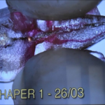
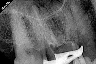
Patient Info:A 65-year-old patient presented with biting pain and a buccal sinus tract (fistula) on the right maxillary first molar (Tooth 16).
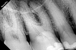
Radiograph:Radiolucency at the apex of the mesiobuccal (MB) root; severe calcification in the pulp chamber and the coronal third of the canals.
Diagnosis:Chronic apical periodontitis with a sinus tract; Severe pulp calcification.
2. Clinical Procedures & Image Sequence
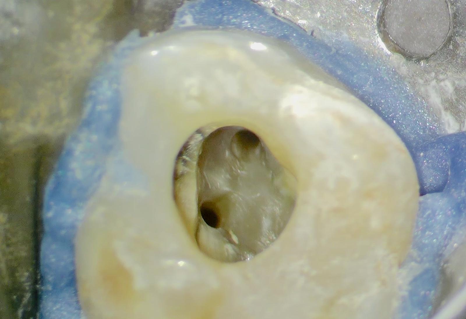
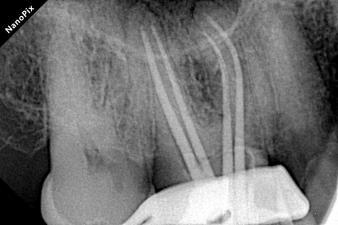
Locating the Hidden Anatomy: Due to heavy calcification, ultrasonic tips were used.
Successfully located the MB1 and the extremely narrow MB2 canal under magnification.
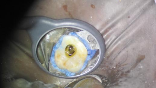
Shaping with Perfection: The canals were shaped using the DENCO Only One File Blue (up to 40.06). The file navigated the extreme curvatures and calcifications flawlessly without any transportation.
Showcasing perfectly tapered and centered canal preparations.
Advanced Disinfection: To ensure the complete eradication of the apical infection, standard irrigation was enhanced by advanced technology.
Use of photodynamic therapy with low-level laser therapy (PDT & LLLT)] applied to maximize bacterial reduction.

Intracanal Medication:Confirming the dense placement of the medication. The sinus tract healed completely within 14 days.
3.Obturation Sequence (Thermoplasticized Technique)
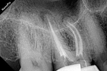
Verifying the precise tug-back fit of the gutta-percha cones.
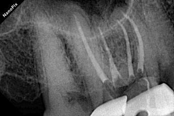
Radiograph of mesio buccal root canals during obturation
Confirming the dense, 3D fill of the entire root canal system.
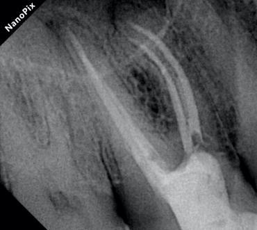
Displaying the excellent final result, particularly highlighting the beautiful flow in the mesiobuccal root canals
4. Clinical Conclusion
In dealing with complex anatomies complicated by severe calcification and periapical lesions, the perfect combination of DENCO APEX-S (for precise WL control) and Only One File Blue (for superior flexibility even at large apical sizes)significantly enhances the safety, predictability, and success rate of complex endodontic treatments.