


Dr. Kais Alshwihdi
Libya
Introduction
Maxillary first premolars are known for their anatomical variability, and cases involving three canals remain relatively uncommon. Successfully managing such complex root canal anatomy requires precision, advanced tools, and careful technique. This case study details the root canal treatment of a maxillary left first premolar (tooth UL4) presenting with irreversible pulpitis and a rare three-canal configuration (buccal-mesial, buccal-distal, and palatal).
The use of the Denco Dual Shaper NiTi rotary file system combined with bioceramic single-cone obturation proved highly effective, turning a challenging case into a predictable success.
Case Diagnosis
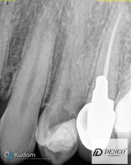
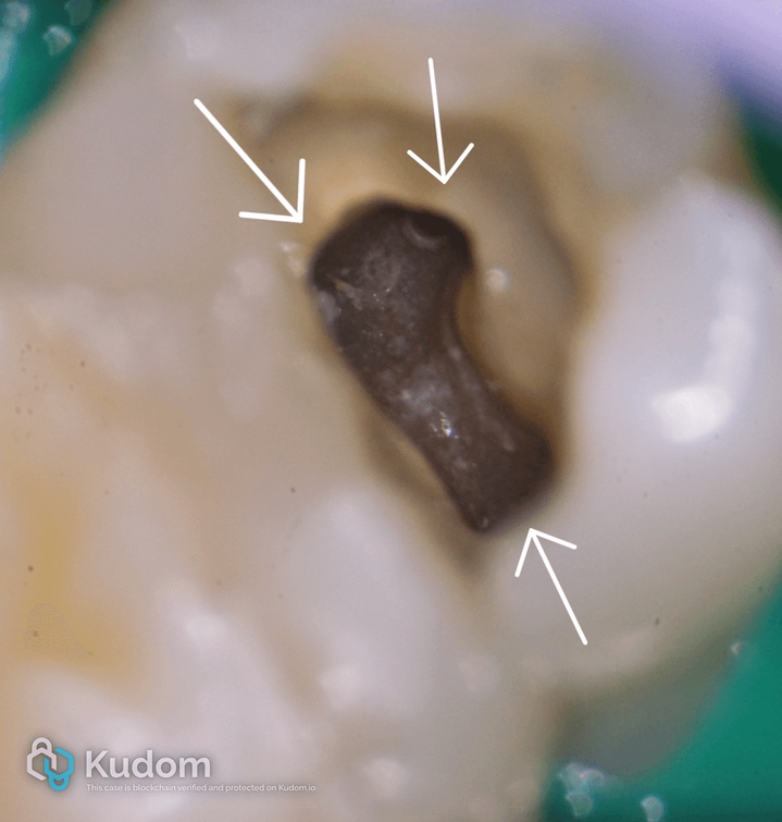
A 32-year-old patient reported spontaneous pain and prolonged cold sensitivity in the upper left first premolar. Clinical examination and radiographic evaluation confirmed:
- Irreversible pulpitis with symptomatic apical periodontitis
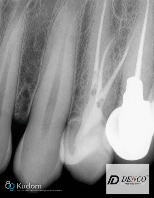
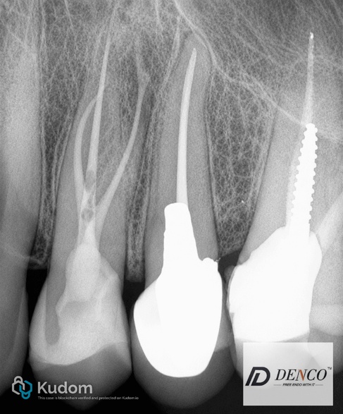
- Complex root canal anatomy: three canals (MB – mesial buccal, DB – distal buccal, and palatal)
Early identification of this complex anatomy was critical to avoid missing canals and ensure complete disinfection.
Treatment Procedure
1. Access and Cleaning
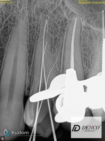
A conservative endodontic access cavity was prepared to preserve as much tooth structure as possible. Under magnification, all three canals were located and negotiated successfully. Working length was determined using an electronic apex locator and confirmed with a radiograph.
2. Shaping with Denco Dual Shaper
Rotary preparation was performed using the Denco Dual Shaper NiTi rotary file system. These files offered several key advantages in this complex premolar case:
- Highly flexible NiTi alloy – excellent performance in curved and narrow canals
- Superior resistance to cyclic fatigue – significantly reducing the risk of file separation
- Ability to preserve original canal anatomy while achieving efficient shaping
- Safe and effective performance in complex premolar anatomies
The Denco Dual Shaper system allowed safe navigation of all three delicate canals without transportation or excessive dentin removal.
3. Irrigation Protocol
Thorough irrigation is essential for successful disinfection, especially in multi-canal systems:
- Sodium hypochlorite (NaOCl) for organic tissue dissolution and antibacterial action
- EDTA to remove the smear layer and open dentinal tubules
- Final rinse with saline
4. Obturation
Obturation was completed in a single visit using the single-cone technique:
- Bioceramic sealer applied for its excellent sealing properties and biocompatibility
- Master gutta-percha cones fitted precisely to working length
- Dense, three-dimensional fill achieved and verified radiographically
Treatment Outcome
The post-operative radiograph demonstrated well-sealed three canals with no voids or gaps. The patient reported complete resolution of symptoms following the procedure.
Key Clinical Results:
- Safe navigation of rare anatomical variations
- Conservative preparation that fully preserved root structure
- Flawless, void-free obturation using bioceramic sealer and single-cone technique
Conclusion
Maxillary first premolars frequently present with anatomical variations that can complicate endodontic treatment. This case highlights how modern rotary instrumentation like the Denco Dual Shaper — with its flexibility, fatigue resistance, and canal-preserving abilities — combined with bioceramic single-cone obturation, delivers efficient, predictable, and successful outcomes even in challenging 3-canal premolar cases.
“Complexity meets simplicity. Dual Shaper turns rare anatomical challenges into predictable, everyday successes.”